
List of publications and related information

Dynamic chromatin association of IκBα is regulated by acetylation and cleavage of histone H4
By |2026-09-01T09:37:56+00:00December 20, 2021|CBBL News, Publications|

IκBs exert principal functions as cytoplasmic inhibitors of NF-kB transcription factors. Additional roles for IκB homologues have been described, including chromatin association and transcriptional regulation. Phosphorylated and SUMOylated IκBα (pS-IκBα) binds to histones H2A and H4 in the stem cell and progenitor cell compartment of skin and intestine, but the mechanisms controlling its recruitment to chromatin are largely unknown. In a recent paper led by Lluís Espinosa at IMIM, the team showed that serine 32–36 phosphorylation of IκBα favors its binding to nucleosomes and demonstrate that p-IκBα association with H4 depends on the acetylation of specific H4 lysine residues. The N-terminal tail of H4 is removed during intestinal cell differentiation by proteolytic cleavage by trypsin or chymotrypsin at residues 17–19, which reduces p-IκBα binding. Inhibition of trypsin and chymotrypsin activity in HT29 cells increases p-IκBα chromatin binding but, paradoxically, impaired goblet cell differentiation, comparable to IκBα deletion. Taken together, the results indicate that dynamic binding of IκBα to chromatin is a requirement for intestinal cell differentiation and provide a molecular basis for the understanding of the restricted nuclear distribution of p-IκBα in specific stem cell compartments.

In summary:

Nuclear IκBα preferentially binds the acetylated N-terminal tail of histone H4 in vivo, specifically in the skin and intestine stem cell compartments. N-terminal cleavage of histone H4 facilitates IκBα dissociation and cellular differentiation.

    • Nuclear IκBα binds the acetylated N-terminal tail of histone H4.
    • The N-terminal tail of histone H4 is lost during cellular differentiation.
  • Histone H4 cleavage is likely produced by Trypsin and Chymotrypsin.
  • Cellular differentiation requires dynamic IκBα chromatin binding and dissociation.

Laura Marruecos, Joan Bertran, Daniel Álvarez-Villanueva, María Carmen Mulero, Yolanda Guillén, Luis G Palma, Martin Floor, Anna Vert, Sara Arce-Gallego, Irene Pecharroman, Laura Batlle, Jordi Villà-Freixa, Gourisankar Ghosh, Anna Bigas, Lluís Espinosa. Dynamic chromatin association of IκBα is regulated by acetylation and cleavage of histone H4. EMBO Reports (2021)22:e52649 2021 https://doi.org/10.15252/embr.202152649
See also comment by Maarten Dhaenens in EMBO Reports.

Selected publication: SBMOpenMM; A Builder of Structure-Based Models for OpenMM

By |2026-09-01T09:38:09+00:00December 20, 2021|CBBL News, PhD, Publications|

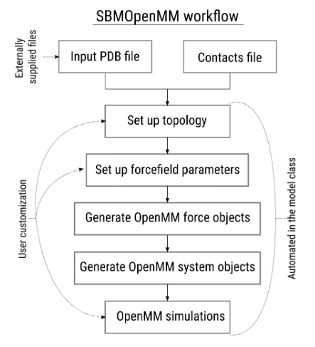
Molecular dynamics (MD) simulations have become a standard tool to correlate the structure and function of biomolecules, and significant advances have been made in the study of proteins and their complexes. A major drawback of conventional MD simulations is the difficulty and cost of obtaining converged results, especially when exploring potential energy surfaces containing considerable energy barriers. This limits the wide use of MD calculations to determine the thermodynamic properties of biomolecular processes. Alternatively, a wide range of Structure-Based Models (SBMs) has been used in the literature to unravel the basic mechanisms of biomolecular dynamics. In a recent article in JCIM, we introduce SBMOpenMM, a Python library to build force fields for SBMs, that uses the OpenMM framework to create and run SBM simulations. The code is flexible, user-friendly, and profits from the high customizability and performance provided by the OpenMM platform.

SBMOpenMM: A Builder of Structure-Based Models for OpenMM

Martin Floor, Kengjie Li, Miquel Estévez-Gay, Luis Agulló, Pau Marc Muñoz-Torres, Jenn K. Hwang, Sílvia Osuna, and Jordi Villà-Freixa
Journal of Chemical Information and Modeling 2021 61 (7), 3166-3171

DOI: 10.1021/acs.jcim.1c00122

Modeling of New Mutations in HFE2 and TFR2 Genes Causing Non HFE-Related Hereditary Hemochromatosis

By |2026-09-01T09:38:15+00:00December 20, 2021|CBBL News, Publications|

Hereditary hemochromatosis (HH) is an iron metabolism disease clinically characterized by excessive iron deposition in parenchymal organs such as liver, heart, pancreas, and joints. It is caused by mutations in at least five different genes. HFE hemochromatosis is the most common type of hemochromatosis, while non-HFE related hemochromatosis are rare cases. Here, we describe six new patients of non-HFE related HH from five different families. Two families (Family 1 and 2) have novel nonsense mutations in the HFE2 gene have novel nonsense mutations (p.Arg63Ter and Asp36ThrfsTer96). In this new collaborative article led by Mayka Sánchez from the Department of Basic Sciences at UIC Barcelona, our lab gave the structural context for the effect of the identified mutations in TFR2 interactions.

Comparative modeling of the TFR2 dimer complexed with TF. (A) TFR2 chains are shown in cyan and pink, while TF chains are shown in green and orange. Dimeric interactions between TFR2 chains, close to Asp680 position (red circle at the α-15 helix, pink chain), are made between residues in the α-18 helix of one TFR2 chain (pink chain) and the loop located between the β-6 and β-7 strands of the other (cyan chain). (B) Sequence alignment of the TFR1 and TFR2 proteins near the TFR2 680 position (green arrow). Numbering is according to the TFR1 sequence, and the secondary structure depiction is based on the TFR1 structure (PDB ID 1CX8). The alignment coloring was produced by the ESPript 3.0 web server [40], where position with red background means absolute sequence conservation. (C–E) Structural context of positions TFR1-Asp648 (C, purple), TFR2-Asp680 (D, pink), and TFR2-Asp680Tyr (E, magenta). Residue names of TFR1/2 appear in white, while residues in TF are in black. Significant interactions are shown with dashed lines.


Hernández, G.; Ferrer-Cortès, X.; Venturi, V.; Musri, M.; Pilquil, M.F.; Torres, P.M.M.; Rodríguez, I.H.; Mínguez, M.À.R.; Kelleher, N.J.; Pelucchi, S.; Piperno, A.; Alberca, E.P.; Ricós, G.G.; Giró, E.C.; Pérez-Montero, S.; Tornador, C.; Villà-Freixa, J.; Sánchez, M. New Mutations in HFE2 and TFR2 Genes Causing Non HFE-Related Hereditary Hemochromatosis. Genes 2021, 12, 1980. https://doi.org/10.3390/genes12121980

New model for CFTR

By |2026-09-01T09:39:12+00:00July 5, 2012|CBBL News, Publications|

The CBBL has completed part of the work of a three years long project on translational bioinformatics in cystic fibrosis (Activa). Our main role in the project was the determination of a model structure for CFTR, the chloride channel whose mutations are in the basis of the disease, as well as being a target for acute watery diarrhea drugs.

The new model, built in collaboration with Nir Ben-Tal’s lab at Tel Aviv University, proposes an open conformation of the channel, oposite to previous models, paving the way for the development of new drugs for both activation and blocking of the channel.

Go to Top