Teeth provide information about the evolutionary pathway of an organism, its biology and habitat. This is the case even of fossilized teeth, since they have perdurable biomineralized structures, as biological apatite. The material that has been selected for this study comprises teeth from modern crocodilian individuals and extinct Cretaceous crocodylomorphs from Lo Hueco site. Microanatomy, histochemistry and crystallographic nature of enamel, dentine and cementum have been characterized by Polarized Light Microscopy, SEM-EDS, Confocal Raman Spectroscopy and SR-µXRD. A focus has been made on dentine lamination. In the fossil sample short-period incremental lines show alternate presence of dentinal tubules that has not been described previously either in living or fossil archosaur. This could be related to influence of environmental circadian rhythms in the abundance, size and/or activity of cells depositing dentine in the day-night cycle. Regarding histochemical and crystallographic compositions, the major and mostly unique phase is HA, but in the case of fossil teeth, a secondary phase identified as hematite appears locally between discontinuities of the material. Incremental lines would not be related to variation in chemical composition and furthermore do not present different HA crystallographic nature (different directions of HA or different crystallite sizes) either. Only small intensity oscillations are observed in the fossil sample by SR-µXRD which are compatible with the alternating abundance of dentinal tubules. Crystallinity differences between modern and fossil material, as crystallite size and presence of CO32- groups could be explained by postdepositional processes.
- Audije-Gil, J., Canillas, M., Barroso-Barcenilla, F., Berrocal-Casero, M., Del Campo, A., González Martín, A., Molera, J., . . . Cambra-Moo, O. (2022). Going deeper into modern and fossil crocodilian tooth microanatomy: what can be inferred of palaeoenvironment and taphonomy from histochemical analyses? Rivista Italiana Di Paleontologia E Stratigrafia, 128(2), 539-557. https://doi.org/10.54103/2039-4942/15607
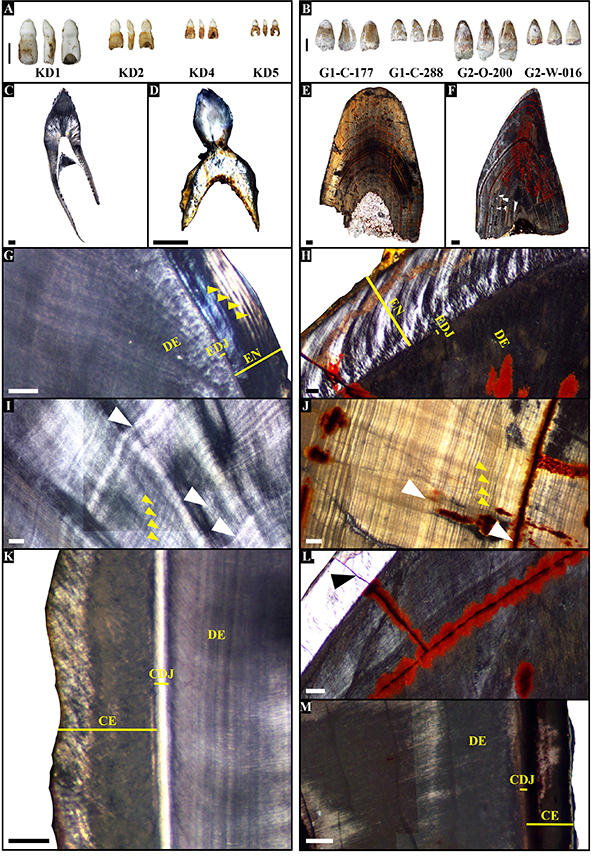
Fig. 1 – Macroscopical and microscopical view (in Polarized Light Microscopy) of the modern (left column) and fossil (right column) crocodilian teeth. A) Modern sample, three views (anterior, right lateral and posterior) per tooth. B) Fossil sample, three views (anterior, right lateral and posterior) per tooth. C-D) Thin section photomontage of modern teeth KD1 (C) and KD4 (D). KD1 shows a replacement tooth in-side still in formation without root (and consequently, with-out cementum) that presents enamel and dentine with their corresponding incremental lines. E-F) Thin section photo-montage of fossil tooth G1-C-177 (E) and G2-W-016 (F). G) Detail of enamel in modern tooth KD1, where lamina-tion parallel to the profile of the EDJ can be observed (yel-low arrows). H) Detail of enamel in fossil tooth G2-W-016, where enamel prisms arrangement can be observed. I) Den-tine in modern tooth KD1, with short-period incremental lines (yellow arrows) and clear-dark intervals representing long-period incremental bands of variable widths (among white arrows). J) Dentine in fossil tooth G1-C-177, with short-period incremental lines (yellow arrows) and clear-dark intervals representing long-period incremental bands of variable widths (among white arrows). Ferruginous mate-rial can be observed between some layers and inside poro-sity. K) Cementum in modern tooth KD1. L) Microfracture (black arrow) in enamel of fossil tooth G2-W-016, through which ferruginous material has infilled. M) Cementum in fossil tooth G2-W-016, partially replaced by ferruginous material. Abbreviations: CDJ = Cementum-dentine junc-tion; CE = Cementum; DE = Dentine; EDJ = Enamel-dentine Junction; EN = Enamel. Scale bars from A-F = 1 mm; scale bars from G-M = 100 μm.
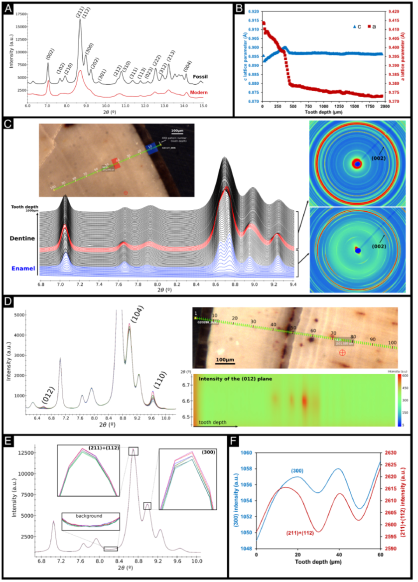
Fig. 4 – Main Synchrotron-radiation X-ray micro-diffraction (SR-μXRD) results of the modern and fossil crocodilian teeth. A) Powder diffraction pattern of the dentine in modern KD1 and fossil G1-C-177 teeth, at 900-micron depth, with the reflection indices of hydroxyapatite (HA) main peaks. A 3000 counts intensity offset has been applied to the fossil tooth data for clarity. B) Evolution of the lattice parameters of HA crystal structure in depth in the fossil tooth G1-C-177. C) Stacking of the powder diffraction patterns along a measurement line from the outside to the inside of the fossil tooth G1-C-177 (100 points spaced by 10 μm between them), through the enamel (blue) and the dentine (black) zones. The red patterns correspond to a zone in the dentine where the HA crystals loose the orientation and experiment a strong reduction of its a lattice parameter. A 2D XRD image is shown for each of the HA states, oriented and non-oriented. D) Stacking of diffraction patterns along a 100 μm zone (10 patterns) with the assignment of the hematite phase peaks identified by Confocal Raman Spectroscopy. The spatial distribution of hematite infilling along a 1000 μm zone for the fossil tooth G2-O-200 according to the intensity of the (012) peak is shown. E) Stacking of powder diffraction patterns through a 60 mm scan (6 patterns) in the fossil tooth G1-C-177, where small intensity variations can be observed on the HA diffraction peaks. F) Variation of the intensity of (211+112) and (300) HA diffraction peaks in depth in the fossil tooth G1-C-177.


